Screening Fresh Oranges With UV:
Study Pinpoints New Value of Detection Tactic
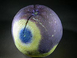
Fresh, deliciously sweet navel oranges, on display at your local supermarket, may have been quickly inspected with ultraviolet (UV) light when they were still at the packinghouse. Usually, the purpose of this special sorting and screening is to see if circular spots—which glow a bright, fluorescent yellow and may be about the size of a quarter or larger—show up on the fruit’s peel.
More often than not, these spots, which scientists refer to as “lesions,” are telltale indicators of the presence of microbes that cause decay, namely Penicillium italicum, responsible for blue mold, or P. digitatum, the culprit behind green mold.
It isn’t the microbes that are fluorescing under the packinghouse UV lamps. Instead, it’s tangeritin, a natural compound in citrus peel oil. When the peel is damaged, such as by decay, tangeritin moves closer to the peel surface, or perhaps seeps out of it, becoming easier for UV to detect.
The characteristic “fluorescence signature” of the decay lesions is easily recognized by packing-line workers who monitor the fruit as it speeds past them on a conveyor belt. All navel oranges that display this distinctive pattern are promptly culled—an established practice that dates back more than 50 years in California citrus packinghouses.
|
|
But studies by plant physiologist Dave Obenland and plant pathologist Joe Smilanick—both with the Agricultural Research Service in Parlier, California—suggest that other, less-studied patterns of fluorescence on navel orange peels may warrant more attention. Fluorescence in the form of specks, smears, smudges, or blotches, for example, may indicate the presence of cuts, punctures, or other peel wounds that might not be visible to the naked eye, yet may pave the way to attack by decay microbes.
To learn more about these less-familiar patterns, the researchers sampled about 5,000 navel oranges over a 2-year period. For the study, navel oranges sampled at two California citrus packinghouses were sorted by fluorescence level—zero, sparse, moderate, or high—noted during UV screening. Next, the oranges were evaluated twice under normal light—not UV. The first time was within 24 hours after UV screening and sorting; the second was after the oranges had been stored at 59°F for 3 weeks.
As expected, fruit with high fluorescence developed further decay and peel-quality problems during storage—but so did many of the oranges that had only moderate fluorescence.
Taken as a whole, the findings suggest that packers might want to expand UV screening to take several fluorescence levels and patterns into account when sorting navel oranges. Many of the patterns that the researchers investigated, such as glowing specks no bigger than the tip of a ballpoint pen, might be quickly and easily detected with modern UV-equipped machine-vision sorters.
The idea of expanding UV use to include more than detection of the classic decay signature is not new. But the Parlier study, though preliminary, is likely the first to present as detailed a look at this approach.
ARS and the grower-sponsored California Citrus Research Board, in Visalia, funded the research.
Obenland, Smilanick, ARS plant pathologist Dennis Margosan at Parlier, and ARS statistician Bruce Mackey at Albany, California, documented the UV findings in a 2010 peer-reviewed article in HortTechnology.—By Marcia Wood, Agricultural Research Service Information Staff.
This research is part of Quality and Utilization of Agricultural Products, an ARS national program (#306) described at www.nps.ars.usda.gov.
David M. Obenland and Joseph L. Smilanick are at the USDA-ARS San Joaquin Valley Agricultural Sciences Center, 9611 S. Riverbend Ave., Parlier, CA 93648; (559) 596-2801 [Obenland], (559) 596-2810 [Smilanick].
"Screening Fresh Oranges With UV: Study Pinpoints New Value of Detection Tactic" was published in the August 2013 issue of Agricultural Research magazine.


