Animal Scientist Finds New
Vistas in Cleft Palate Research
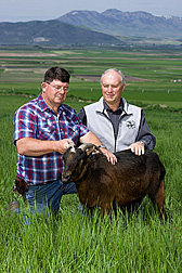
Agricultural Research Service (ARS) animal scientist Kip Panter began by studying why some cows gave birth to offspring crippled by “crooked calf syndrome,” or CCS. Now he has the answer—and he’s using his findings to help doctors develop protocols for repairing a common birth defect in humans.
How did an agricultural scientist who spends much of his time studying livestock production end up partnering with pediatric plastic surgeon Jeffrey Weinzweig? “Research is sometimes serendipitous,” Panter says simply. “All mammals are related to some degree. So sometimes our work for improving livestock health can also have big payoffs in human health.”
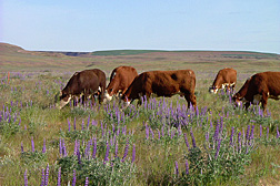
Much of Panter’s research has its roots in the 1960s. This is when scientists first realized that pregnant cows that grazed on lupines ran a significantly higher risk of giving birth to calves affected by CCS. These calves are usually born alive at full term, but they can have a number of birth defects, including skeletal deformities and cleft palate.
More often than not, calves with CCS don’t survive. It is possible to save less severely affected animals, but they often need assisted deliveries or other interventions that can require up to 60 percent more care from a veterinarian. Even after these efforts, the calves remain unmarketable.
The toll can be devastating to livestock producers and cattle alike. For instance, in 1992, an Oregon rancher lost 56 percent of his calves to CCS. In 1997, more than 650 calves were lost to CCS in east-central Washington, and some ranchers lost all their calves.
The link between lupine consumption and CCS exists because some lupine varieties contain concentrations of alkaloid toxins that ebb and peak throughout the life of the plant. Livestock, including pregnant cattle, usually begin to graze lupines in early July—a schedule that overlaps an interlude during gestation when fetal calves begin the physical activity essential for normal development.
This activity starts with fetal head and neck extensions and is followed by other vigorous and frequent physical movements. But during this time, alkaloids in the lupines can cross the placental barrier and temporarily induce fetal immobility.
Working at the ARS Poisonous Plant Research Laboratory in Logan, Utah, chemist Richard Keeler (now retired) started to identify alkaloids in lupines that contributed to the onset of CCS.
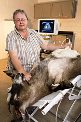
After several rounds of testing with pregnant cattle, Keeler determined the alkaloid anagyrine was the primary substance in lupines that affects cattle. At this time, Panter joined the team and used ultrasound to confirm that when pregnant Spanish goats consumed the toxic alkaloids, fetal immobility resulted.
Then the team conducted ultrasounds on pregnant cows that had consumed the toxic plants at some point throughout the crucial window in fetal development. The scientists identified a direct relationship between lupine consumption, reduced fetal movement, and severity of skeletal defects in the offspring.
“We knew that the malformations develop because some lupine alkaloids cross the placental barrier and have a neuromuscular blocking effect,” Panter explains. “But these tests showed us that it’s the reduction in fetal movement that in turn impairs normal skeletal development.”
Panter also figured out the link between lupine consumption and the development of cleft palate.
“In the developing goat and cow fetus, the tongue is positioned between the palatal shelves until the fetus begins to move. The palate typically closes around 38 days after conception in goats and around 50 days in cows. But if the fetus isn’t physically active during that time, the position of the tongue prevents the closure, and a cleft palate results,” Panter explains.
Keeler and Panter also conducted a round of research to see whether the alkaloid anabasine in wild tree tobacco (Nicotiana glauca)—a plant that is also toxic to livestock—had similar effects on Spanish goats. Panter fed the plant to three pregnant goats from 35 to 41 days after they conceived.
Ultrasounds confirmed that throughout their exposure to anabasine, the fetal goats remained motionless. When the kids were born, Panter expected that they would all have cleft palates—and they did. But because the fetuses were exposed to the alkaloid for a brief specific time, they only developed cleft palates. More severe skeletal defects did not result.
The Animal Scientist and the Plastic Surgeon
In 1994, Panter was contacted by Weinzweig, who was developing prenatal surgical techniques to repair cleft palates in humans. The second most common birth defect in the United States, clefting anomalies—cleft lips or palates or both—affect about 1 in every 700 people.
A cleft palate is not life threatening. A range of surgical procedures, which typically start before a baby is a year old and often continue into adulthood, can be used to reconstruct much of the palatal anatomy disrupted by clefting. But even the most gifted plastic surgeons can find successful palate repair a challenge.
So Weinzweig wanted to explore what is called the “privileged” window of fetal healing. Before birth, damaged fetal tissue can often heal without any scarring. Weinzweig wanted to see if prenatal, or in utero, cleft palate repair could result in a normal functional palate by facilitating development of the necessary structural components—muscle, bone, and the mucous membranes and fibrous connective tissue that cover the hard palate.
Weinzweig needed a suitable animal model for experimental surgery. He thought that Panter’s findings with the goats might hold the key to that part of the puzzle.
“I thought Dr. Panter’s research was brilliant,” Weinzweig says. “It seemed completely possible that we could use this work to define a model for characterizing the mechanisms in cleft palate development and repair.”
Other researchers had used sheep models to investigate prenatal cleft palate repairs of surgically created clefts. But their results were not as applicable to the human clefting anomaly as the congenital model Panter and Weinzweig began to develop with the Spanish goats. In addition, the sheep model did not provide reliable or reproducible results.
So Panter and Weinzweig ran another trial that compared clefting between the two animals, which confirmed that goats were the best candidates for the research. Then they fed six pregnant goats wild tree tobacco during the narrowly defined fetal period and confirmed via ultrasound that fetal activity temporarily ceased during exposure to the toxin.
A New Model for Prenatal Surgery
Over the next decade, Weinzweig and half a dozen surgical residents performed prenatal cleft palate repairs on dozens of fetal goats that had been exposed in utero to the wild tree tobacco toxin. These procedures were first conducted at Weinzweig’s lab at Brown University’s Alpert Medical School in Providence, Rhode Island. But after 2003, the surgeons traveled to Logan, where the newly renovated ARS laboratory provided state-of-the-art facilities for the procedures.
All the pregnant goats and all the fetuses survived the surgeries without complications. Throughout their research, the team maintained a meticulous series of tissue and X-ray studies from the goats that had undergone prenatal repairs and from groups of control animals that were not clefted. They began collecting data soon after the goats were born and continued tracking their progress until the goats were 6 months old.
The studies showed that after birth, the muscle and mucosal structures of the repaired palates were virtually indistinguishable from the palates of goats that had never developed clefting defects. The procedures also resulted in substantial repair of clefted bony structures, although these results didn’t match the near-flawless response of the muscle and mucosal tissues. Just as important, the prenatal surgery completely restored palatal function, which allowed the goats to nurse as soon as they were born and to vocalize without impediments.
The in utero-repaired palates had none of the scarring typically associated with postnatal cleft palate repairs. But, despite the successful healing, the kids still had maxillas—or upper jaws—that were disproportionately smaller than their lower jaws. Until recently, doctors believed that this size difference in clefted humans resulted from scarring that developed after palate repair and restricted proper growth.
“We’ve confirmed that even though the size difference can be exacerbated by surgery, the difference is primarily due to the clefting anomaly itself,” says Weinzweig, who now chairs the Department of Plastic and Reconstructive Surgery at Lahey Clinic Medical Center in Burlington, Massachusetts. “When one of my colleagues heard about this, he said, ‘This is wonderful! For the first time, we have the data to end the controversy over how this anomaly develops.’”
Though confident that the prenatal cleft repair technique could be effective for humans, Weinzweig cautions that it would not even be attempted until proven protocols are in place to protect the safety of the mother and the fetus. “Prenatal surgery in humans is currently performed only for a few life-threatening conditions, such as diaphragmatic hernia, hydronephrosis, and spina bifida,” he notes.
Panter is delighted that Weinzweig’s techniques have been so successful to date and that so many surgeons-in-training have been part of the project. At the same time, his main focus remains on supporting livestock producers.
“We’re still fine-tuning the prenatal cleft palate repair. We’re also developing new supports for postnatal palate surgeries,” Panter says. “But CCS can take a heavy toll on cattle, and producers pay a high price—both economically and emotionally—when their animals are affected. So we’ll keep working with ranchers to develop and implement effective grazing-management plans that protect cattle herds from CCS.”—By Ann Perry, Agricultural Research Service Information Staff.
This research is part of Pasture, Forage, and Range Land Systems, an ARS national program (#215) described on the World Wide Web at www.nps.ars.usda.gov.
Kip E. Panter is with the USDA-ARS Poisonous Plant Research Laboratory, 1150 East 1400 North, Logan, UT 84341; phone (435) 752-2941 (ext. 1123), fax (435) 753-5681.
"Animal Scientist Finds New Vistas in Cleft Palate Research" was published in the August 2009 issue of Agricultural Research magazine.


