United States Department of Agriculture
AgResearch Magazine


Where's the Fat?
|
|
No one could call Al Mitchell's Sundays relaxing.
First, there's the task of negotiating a truckload of a dozen or so hefty hogs through downtown Washington, D.C., traffic. Then, Mitchell gets to help lift each of the hogs onto a high table—twice—at their destination, before heading home with his squealing load at the end of a long, long day.
Mitchell's no moonlighting hog farmer; he's an animal scientist with USDA's Agricultural Research Service.
An even bigger surprise is the hogs' destination on their Sunday jaunts:
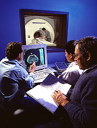
Howard University, where they undergo magnetic resonance imaging (MRI)—the same high-tech procedure medical doctors use to detect internal disorders such as brain tumors in humans.
Since 1985, Mitchell has collaborated with Paul Wang of Howard University on using MRI's to analyze body composition of research hogs while they're still on the hoof.
The sharp images of the MRI's let scientists see how a particular swine breeding line looks—in terms of fat-to-lean ratio, or how a particular diet is affecting an animal's body—and still use that animal for further research.
"Before MRI's, there really was no accurate way of measuring the body composition of a live animal," explains Mitchell, who is with the ARS Growth Biology Laboratory at Beltsville, Maryland. "The MRI provides a lot of information you can't get with other techniques, such as the amount of fat on the animal, the distribution of that fat, or the volume of muscle in specific muscle groups.
|
|
Magnetic resonance involves placing a specimen—such as a 90-kilogram (200-pound) porker—inside a device with a strong magnetic field, which causes certain naturally magnetized atoms in the specimen to orient themselves in the field, much as a compass needle lines up with the poles in the Earth's magnetic field.
While the atoms are lined up, a short pulse of radio waves is emitted to give them a little push. When that pulse ends, the atoms bounce back into alignment, giving off a faint radio signal that's picked up by the magnetic resonance device. These signals are transmitted to a computer for analysis and converted into a visual image.
"Since this is not a destructive technique, we can follow changes in the animal's body composition as it grows," notes Mitchell.
While the MRI's are as clear as a pork chop on a plate, obtaining them is no picnic. Each pig is anesthetized, then lifted onto the magnetic resonance device's table. Once a pig is in place, a body section up to 40 centimeters long is scanned at increments of one centimeter; then the pig must be repositioned on the table for another scan. At an average of nearly 13 minutes per scan and four scans per pig, a 15-pig day seems interminable.
Mitchell's studies have focused on the impact of diet on pigs, while colleague Armin M. Scholz has used the MRI's to study body type of pigs susceptible to porcine stress syndrome. A visiting scientist from Humboldt University in Berlin, Germany, Scholz says PSS-positive pigs are leaner.
|
|
"In my diet study, I scanned 601 pigs, starting at 20 kilograms [about 44 pounds] each, and I followed them to 60 kilograms [about 132 pounds]," Mitchell recalls. "I slaughtered animals from each weight group and did chemical analyses of the tissue to determine if the MRI was presenting an accurate picture of fat and lean. There was a good match between the chemical analyses and what I saw on the MRI's."
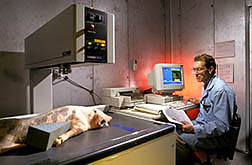
Mitchell can also check body composition closer to home with a Dual-Energy X-ray Absorptiometry (DEXA) unit next to his Beltsville lab. Unlike the MRI, which presents images of cross-sectional "slices" of the pig's body, the DEXA is a whole-body scan without the distinct internal views.
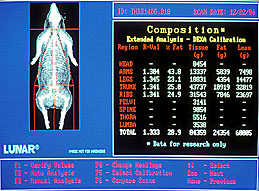
DEXA uses two different energy-levels of X-rays to differentiate between lean and fat, providing percentages and grams of fat and lean tissue throughout the whole body or in a particular area, such as a front or rear leg.
"This system is often used for human body composition studies, but this is the first time it's been used extensively for farm animals," says Mitchell. "DEXA images are not as precise as those in an MRI, but the unit's less expensive and easier to work with technically. Plus, you can get an image of a 90-kilogram pig in 35 minutes.
"These techniques would not be used by the farmer—the equipment is still too expensive," Mitchell says. "But as research tools, they provide a way for us to evaluate an animal without harming it. You don't want to have to slaughter a valuable animal just to see how it's growing." — By Sandy Miller Hays, ARS.
Al D. Mitchell is with the USDA-ARS Growth Biology Laboratory, 10300 Baltimore Avenue, Beltsville, MD 20705-2350; phone (301) 504-8868, fax (301) 504-8623.
"Where's the Fat?" was published in the July 1995 issue of Agricultural Research magazine.