United States Department of Agriculture
AgResearch Magazine


Anatomy of a Snowflake
|
|
Powerful microscope reveals structural details to help predict next summer's water supply.
A snowfall can bring mixed emotions to many people. It can mean a harried drive home during rush hour traffic or the promise of a weekend of skiing.
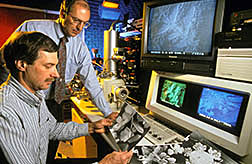
But ARS microscopists William P. Wergin and Eric F. Erbe view snowfalls as welcome opportunities to photograph snowflakes using a scanning electron microscope (SEM).
Images from this instrument capture surreal pictures of microthin needles, hollow columns, hexagonal plates, bullets, and other crystalline formations.
Last winter, the two ARS scientists explored the SEM's potential to yield new clues about the water that's stored as snow crystals in mountain snowpacks. As spring temperatures rise, water from the snowpacks replenishes reservoirs vital to the irrigated agriculture of western states.
The amount of water in a mountain snowpack can vary greatly, depending on the shape and size of its snow crystals, says Wergin, who heads ARS' Electron Microscopy Laboratory located at the Beltsville (Maryland) Agricultural Research Center.
There, Wergin, Erbe, and colleagues devise new techniques and technologies in electron microscope imaging for many applications. For example, their work enables plant physiologists to observe how pollutants, drought, or other environmental stresses affect such processes as cellular growth and development in crop plants.
|
|
Wergin says they first struck upon the idea of photographing the snow-flakes on December 28, 1993. That's when freshly fallen snow afforded them a ready source of material for testing a new cryosystem.
The cryosystem is part of a preparation process in which tissue samples from plants or other specimens are deep-frozen in liquid nitrogen and then coated in a fine layer of metal, before SEM imaging.
"Anything observed in the SEM must be conductive to electrons," Wergin says. "We accomplish that by putting a very thin film of metal such as platinum or gold directly onto the specimen."
Originally, he says, they had intended to test the new cryosystem using a plant specimen or unsuspecting spider or cockroach caught lurking about the lab. But a recent extermination had rid the lab of bugs. And a peek outside confirmed that winter's arrival had killed off the dandelions, thistles, and other plant life growing along the lab's perimeter.
Snow, they thought, would prove an interesting challenge.
Intrigued by the possibilities, the two researchers set about figuring how to collect snow samples from the roofs and bumpers of their cars.
After some trial and error, they settled on the use of a flat copper holder, about an inch square and coated with a glue-like substance known as Tissue Tek. That arrangement enabled them to successfully capture a sampling of snowflakes.
Next, they proceeded to deep-freeze the snow crystals in liquid nitrogen at -320° F, a temperature that will shatter a dropped rubber ball as if it were a crystal goblet. The researchers then coated the snow-flakes with platinum using an instrument called a sputter coater.
That accomplished, they placed the still frozen, coated snow crystals on a cold stage in the microscope.
There, the SEM passed a beam of electrons over the crystals' platinum surface, shaking loose their secondary electrons. These were then recorded by a special detector in the microscope and reproduced as black-and-white SEM snowflake images.
"To our knowledge," says Wergin, "that was the first time snowflakes had been photographed using an SEM."
"They were so intriguing," Erbe recalls of the first set of SEM images. He initially anticipated that they'd reveal something more like the snowflakes portrayed on Christmas cards and ski resort signs.
"What we typically call snowflakes," Erbe says, "are each made up of between two and several hundred snow crystals."
At magnifications of 10,000 to 20,000—well beyond those achieved with conventional light microscopes—the SEM images allow detailed observation of the snow crystals and the manner by which they're configured, broken, and reformed during their descent by a combination of temperature, humidity, and other atmospheric conditions.
"All the existing terminology describing snow crystal shape is based on photographs taken with light microscopes or 35-mm cameras at very low resolutions," Wergin says. "But with SEM images, researchers might discover entirely new crystal structures requiring a new set of descriptive terms."
|
|
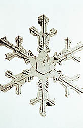
Using stereoscopic techniques, he and Erbe have also produced three-dimensional photos of snow crystals, enhancing the angles from which their structures can be observed. "All snowflakes are based on a hexagonal form, due to the molecular structure of water," Wergin notes.
Better Snowpack Measurements
From this winter's studies, the two researchers hope to exploit the SEM technology to furnish hydrologists with better measurements on snow crystal size and shape, two of the features that dictate a snowpack's water content. Their endeavor could add a valuable new source of data to computer models hydrologists use to forecast snowmelt.
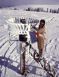
In states like California, Colorado, and New Mexico, spring forecasts tell farmers, ranchers, electrical power plant operators, and others how much water will be available during the summer for crop irrigation, livestock, electricity, and other needs, such as for drinking.
"In the western United States, as much as 75 percent of the water you get in streams comes from snow-melt," says hydrologist Albert Rango, who heads the ARS Hydrology Laboratory in Beltsville.
"The snowmelt runoff season starts around April 1st and may not end until sometime in August," says Rango, whose research includes running ARS' Snowmelt Runoff Model (SRM) to study and forecast snowmelt in mountain regions of both the western and eastern United States.
"For agriculture, it's important to predict water availability relatively early in the planting season, so farmers can know which types of crops will grow to their full yield.
Those same forecasts are also important for other applications, like scheduling the best time to generate hydroelectric power."
Rango says the SEM technology and snow crystal sampling techniques developed by Wergin and Erbe might also be useful in his research on passive microwave sensing, a satellite capability that could bolster the forecasting ability of SRM and other snowmelt models.
In cooperative research, Rango and scientists at the NASA facility in Greenbelt, Maryland, are studying the use of data from passive microwaves naturally emitted from the earth to better estimate the snow water equivalent, or water yield, of snowpacks across broad regions of mountain terrain.
Their research aims to furnish snowmelt models such as SRM with more timely and broad-ranging data about the internal characteristics of snowpacks. This is a feat not easily achieved using data from mountain research sites where automated stations and other sampling devices collect readings on the precipitation, snow depth, temperature, and other conditions in the immediate region.
Rango says one hurdle they've encountered in passive microwave sensing is its inability to distinguish among the varying sizes of snow crystals in a snowpack. In addition to atmospheric conditions encountered during its descent, a snow crystal's size is determined by its depth within a snowpack and by temperature and moisture differences inside and outside the snowpack.
Passive microwave techniques compensate for this lack of precise information by using an average snow crystal size in computer algorithms that estimate the snow water equivalent.
Unfortunately, says Rango, algorithms produce poor water equivalent estimates if the sizes of snow crystals in a snowpack differ significantly from the algorithm's average values.
"What we want to do is find a way to solve this size problem in our models," Rango says. "This may mean finding a better way to estimate not only crystal size, but also crystal shape so the algorithms can produce better results."
He says that this might be accomplished using measurements drawn from SEM photos of snow crystals collected from mountain research sites. To allow for that possibility, Wergin and Erbe adapted special containers for transporting frozen specimens that could also be used to store these snow crystal samples.
Equipped with the containers, a researcher would be able to visit a site, collect snow crystal samples from a snowpit, and preserve them using techniques developed by the two microscopists.
Their techniques would call for the researcher to dip the snow crystal samples into a portable canister of liquid nitrogen, remove the samples, then position them inside special clips of a precooled specimen container. "You can't ship liquid nitrogen on a plane," Wergin says, "but you can use it to bring down the temperature of these containers for about 3 days. That is more than enough time to transport a sample from most areas of the country."
On its arrival at the lab, the snow crystal sample could be coated in platinum and run through the SEM.
Wergin says a calibration device would enable scientists to measure the size, diameter, weight, and other features of the snow crystals with little risk of melting or sublimation, the natural evaporative processes that can confound a researcher's onsite measurements.
And with the future installation of new x-ray equipment, "the SEM would enable us to do an elemental analysis of a snowflake sample," Wergin says. "This technique would allow us to quantify the amount of pollutants, such as sulfur, in falling snow." -- By Jan Suszkiw, ARS.
William P. Wergin and Eric F. Erbe are with the USDA-ARS Beltsville Agricultural Research Center, 10300 Baltimore Ave., Beltsville, MD 20705-2350; phone (301) 504-8046, fax (301) 504-8923.
Albert Rango is with the USDA-ARS Range Management Research, 2995 Knox St., Las Cruces, NM, 88003; phone (505) 646-2120, fax (505) 646-5889.
"Anatomy of a Snowflake" was published in the April 1995 issue of Agricultural Research magazine.